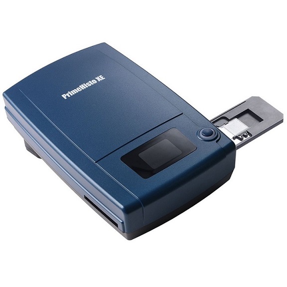
PrimeHisto XE
▪ Capture crisp details with 10,000 dpi true-color linear CCD
▪ Digitalize whole slides for efficient microscope navigation
▪ Empower histology researchers, clinical labs, and educational institutions
▪ Analyze minerals and materials using the rotatable polarizer
▪ Full compatibility with Windows and Mac operating systems

A manual microscope slide scanning scanner offers exceptional quality, best support for microscope use.
- Manual microscope slide scanning - PrimeHisto XE is designed specifically for medical/pathological tissue sample observations.
- Rotatable Polarizer – Built-in design supporting bright field and polarized light scanning for mineral and material analysis.
- With Holders - The slide holder holds 2 tissue slides and guarantees consistent focus quality for standard slide thicknesses ranging from 0.8mm to 1.5mm.
- Pacific Image Exclusive Software Included - HistoView ensures objective scan analysis.
PrimeHisto XE ensures true color reproduction using a True Color Linear CCD. The self-adaptive stage maintains optimal focus across various slide thicknesses, while the 10,000 dpi resolution captures every detail for histology, biology, and mineralogy analysis. This scanner provides a wide field of view with no edge distortion, making it a cost-effective solution for digital navigation.
10,000 dpi Resolution
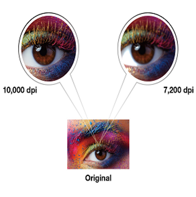
With the advanced resolution, PrimeHisto XE is capable of capturing and digitizing a sharp and vivid image from tissue slides.
Durable Tissue Slide Holder
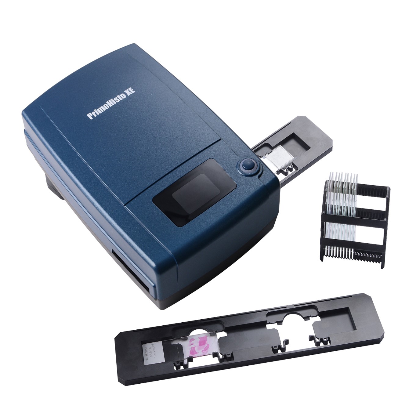
Users can manually insert tissue slide holders made of durable plastic with pressure strips to ensure ensures consistent focus quality across various slide thicknesses.
True Color
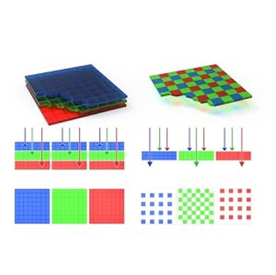
Different from DSLR or CMOS scanners using area sensors with Bayer Patterns to simulate RGB, it is a true RGB linear array CCD, which provides high quality scanning at spatial and color intensity resolution.
Digital Navigation Map

PrimeHisto XE can observe a range of about 10 μm to 1 cm. This capability can effectively assist in locating areas of interest when using a microscope to examine specimens.
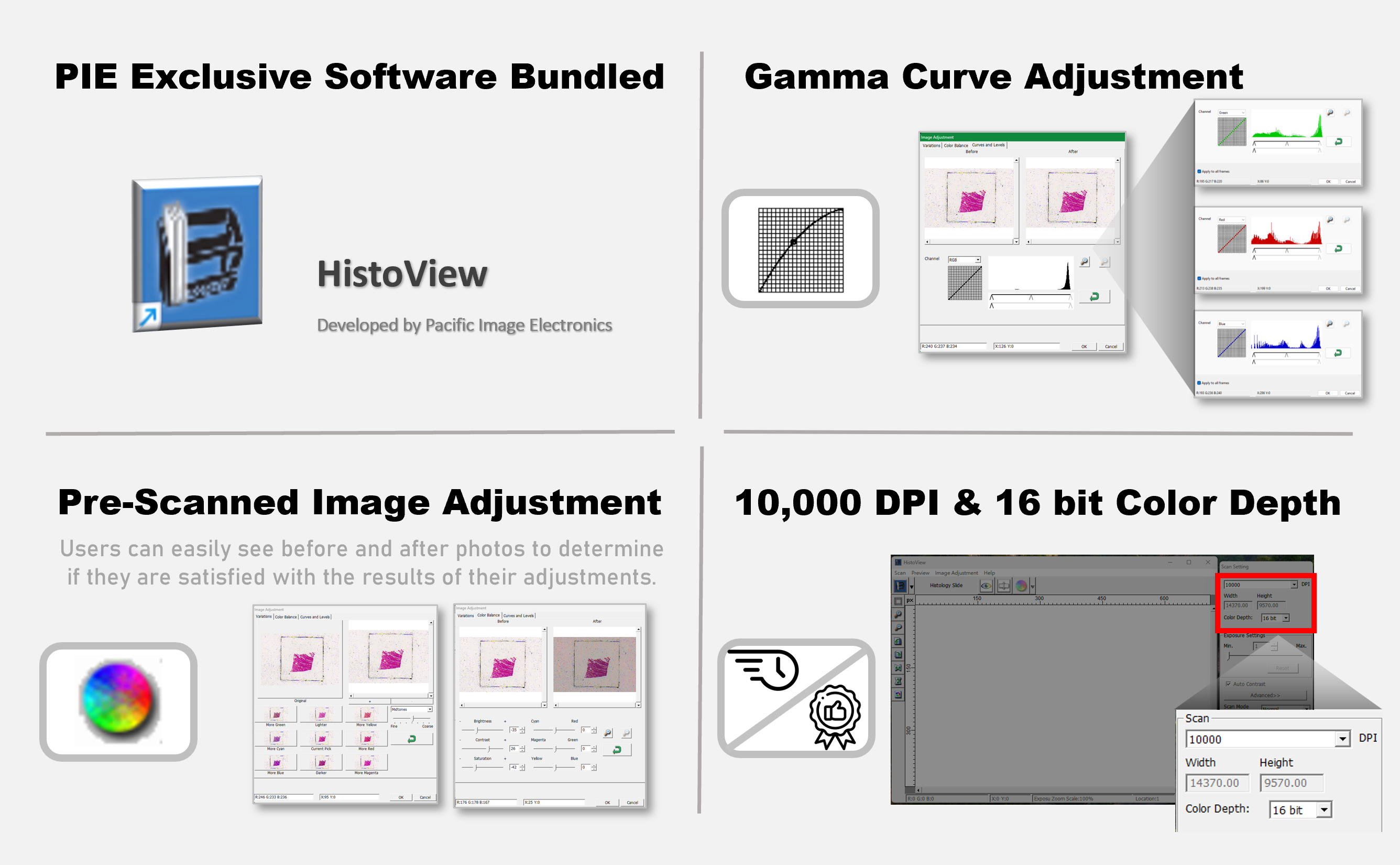
HistoView - Pacific Image Electronics Exclusive
- Gamma Curve Adjustment - Enhances brightness and contrast for vibrant visuals.
- Image Adjustment - Customize brightness, contrast, and saturation.
- 10,000 DPI & 16 Bit Color Depth - Achieve precision in imaging and design projects.
PrimeHisto XE Specifications
Scanning Media:
Microscope Slide
Microscope Slide
Resolution:
10,000 DPI
10,000 DPI
Dynamic Range:
3.9
3.9
Light Source:
Light Transmission White LED
Light Transmission White LED
Sensor:
Linear Array Color CCD
Linear Array Color CCD
Data Conversion:
48 Bits/px (color mode), 16 Bit/px (grayscale mode)
48 Bits/px (color mode), 16 Bit/px (grayscale mode)
Scanning Area:
24.3mm x 36.5mm (H x W)
24.3mm x 36.5mm (H x W)
Image File Format:
TIFF, BMP, JPEG
TIFF, BMP, JPEG
Power Supply:
AC 100-240V; 50/60 Hz Output: 12V DC / 1.5A
AC 100-240V; 50/60 Hz Output: 12V DC / 1.5A
Dimensions:
275 (L) x 167 (W) x 80 (H) mm
275 (L) x 167 (W) x 80 (H) mm
Net Weight:
4.63lb (2.1kg)
4.63lb (2.1kg)
OS:
Windows 7/8/10/11, Mac OS 10.13 (minimum)
Windows 7/8/10/11, Mac OS 10.13 (minimum)
Hardware Requirements:
RAM: 4GB (8GB or more recommended)
RAM: 4GB (8GB or more recommended)
| Scanning Media | Microscope Slide |
| Resolution | 10,000 DPI |
| Dynamic Range | 3.9 |
| Advanced Features | Manual Microscope Slide Scanning |
| User friendly interface |
• One Button Scan • Viewing window |
| Light Source | Light Transmission white LED |
| Sensor | Linear Array Color CCD |
| Data Conversion | 48 Bits per pixel (color mode) 16 Bits per pixel (grayscale mode) |
| Image File Format | JPG, TIF |
| Scanning Area | 24.3mm x 36.5mm (H x W) |
| Interface | USB 2.0 |
| Power Supply |
Image: AC 100-240V; 50/60 Hz Output: 12V DC / 1.5A |
| Accessories | • Tissue Slide Holder • Wall-Mount Adapter • USB 2.0 Cable • Quick Installation Guide |
| Dimensions | 275 (L) x 167 (W) x 80 (H) mm (10.8 x 6.57 x 3.15 inch) |
| Net Weight | 4.63lb (2.1kg) |
| OS |
Windows 7/8/10/11 Mac OS 10.13 (minimum) |
| Hardware Requirements |
Windows Mac |
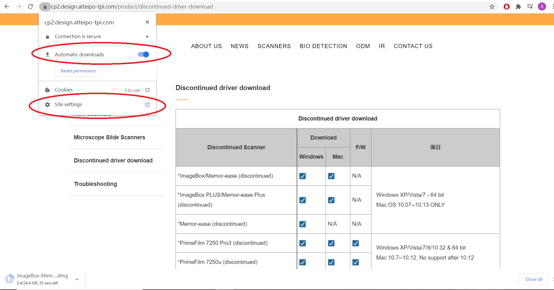
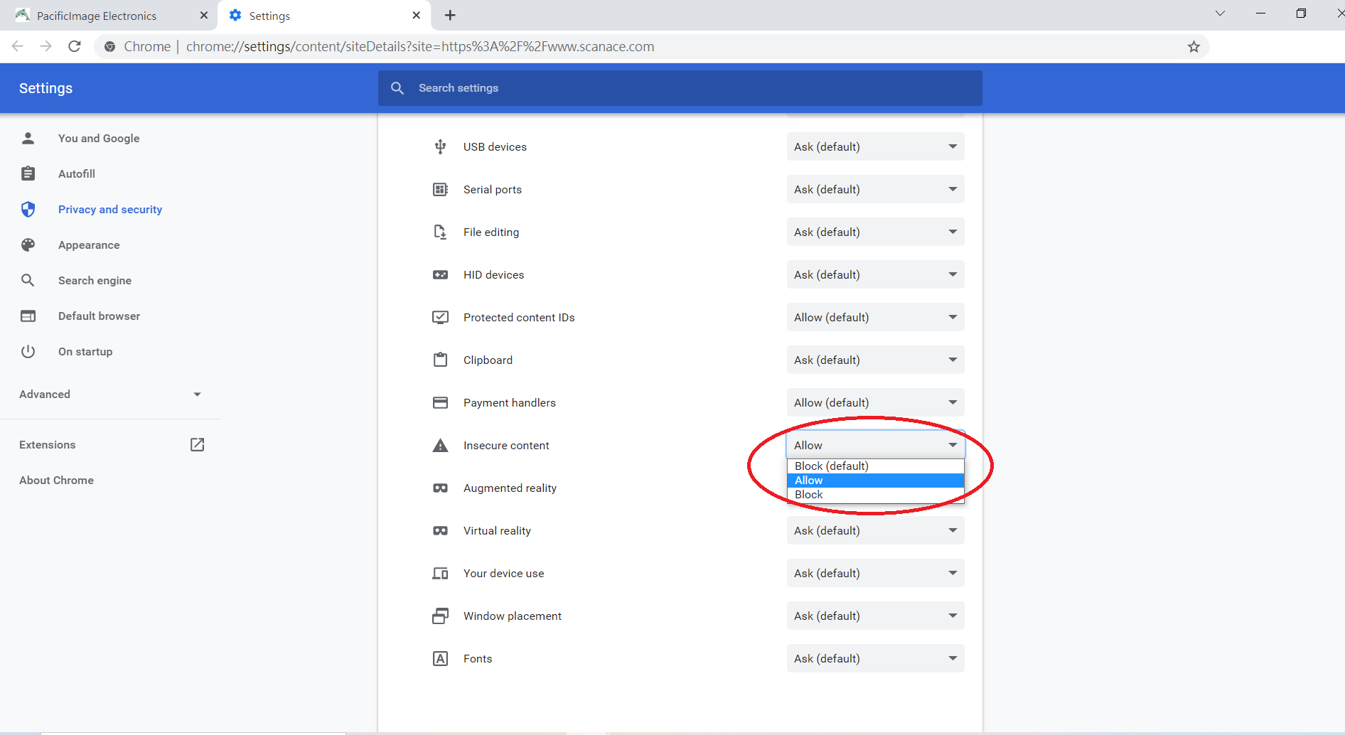
If you are using Google Chrome, kindly refer to the following steps before downloading:
1.Open "Automatic downloads" in the upper left corner on the page of Chrome
2. Click "Site settings"
3. Go to "Insecure content" row and chose "Allow"
If there you are using IE, please refer to the following link:
How to Download software from Scanace Website.pdf
Open a Mac app from an unidentified developer – Apple Support (UK)
Driver for Mac OS 10.13 or higher
Driver for Windows10 32 & 64 bit